Spleen - structure shape, topography, Blood vessels, Nerves innervation
Spleen - size, structure and shape, topographic anatomy, Function, Blood vessels, Nerves, and innervation
Spleen. The size of the spleen. The structure of the spleen. Spleen shape
The spleen, lien (Greek splen), is a richly vascularized lymphoid organ. In the spleen, the circulatory system is closely related to the lymphoid tissue, due to which the blood here is enriched with a fresh supply of leukocytes developing in the spleen.
In addition, the blood passing through the spleen is released due to the phagocytic activity of spleen macrophages from obsolete red blood cells ("graveyard" of erythrocytes) and from pathogenic microbes, suspended foreign particles that have entered the bloodstream, etc.
The size of the spleen, due to the richness of the vessels, can vary quite significantly in the same individual, depending on the greater or lesser filling of the vessels with blood. On average, the spleen is 12 cm long, 8 cm wide, 3-4 cm thick, and weighs about 170 g (100-200 g).
During digestion, the spleen is enlarged. The color of the spleen on the surface is dark red with a purple tint.
The shape of the spleen is compared to a coffee bean. In the spleen, two surfaces are distinguished (facies diaphragmatica and facies visceralis) , two edges (upper and lower) and two ends (anterior and posterior). The most extensive and laterally facing facies diaphragmatica is convex, it is adjacent to the diaphragm.
On the visceral concave surface, in the area adjacent to the stomach (facies gastrica) , there is a longitudinal groove, hilus lienis - the gate through which the vessels and nerves enter the spleen. Behind the facies gastrica there is a longitudinally located flat area, this is the facies renalis , since here the spleen is in contact with the left adrenal gland and kidney.
Near the posterior end of the spleen, the place of contact of the spleen with the colon and lig is noticeable. phrenicocolicum; it is facies colica.
The structure of the spleen. In addition to the serous cover, the spleen has its own connective tissue capsule, tunica fibrosa , with an admixture of elastic and non-striated muscle fibers. The capsule continues into the thickness of the organ in the form of crossbars, forming the skeleton of the spleen, dividing it into separate sections. Here between the trabeculae is the spleen pulp, pulpa lienis . The pulp is dark red in color.
On a fresh cut in the pulp, lighter colored nodules are visible - folliculi lymphatici lienales . They are round or oval lymphoid formations, about 0.36 mm in diameter, sitting on the walls of arterial branches. The pulp consists of reticular tissue, the loops of which are filled with various cellular elements, lymphocytes and leukocytes, red blood cells, most of which are already decaying, with pigment grains.
Spleen Anatomy Video Lesson
Spleen topography. Spleen function
The spleen is located in the left hypochondrium at the level from IX to XI ribs, its longitudinal axis is directed from top to bottom and outward and somewhat forward almost parallel to the lower ribs in their posterior sections.
There is a high position of the spleen when its anterior pole reaches the VIII rib (observed with a brachymorphic physique), and low, when the anterior pole lies below the IX rib (observed with a dolichomorphic physique).
The peritoneum, growing together with the capsule of the spleen, covers it from all sides, with the exception of the gate, where it bends over the vessels and passes to the stomach, forming a lig. gastrolienale. From the gate of the spleen to the diaphragm near the entrance of the esophagus, a fold of the peritoneum (sometimes absent) stretches - lig. phrenicolienale.
Also, lig. phrenicocolicum, stretched between the colon tranusversum and the lateral wall of the abdomen, in the region of the left XI rib, forms a kind of pocket for the spleen, which rests against this ligament with its lower end.
Function. The lymphoid tissue of the spleen contains lymphocytes involved in immunological reactions . In the pulp, the death of a part of the formed elements of blood occurs, the life of which has expired.
The iron of hemoglobin from the destroyed erythrocytes is sent through the veins to the liver, where it serves as a material for the synthesis of bile pigments.
Spleen percussion video
Spleen vessels. Spleen blood supply. Nerves and innervation of the spleen
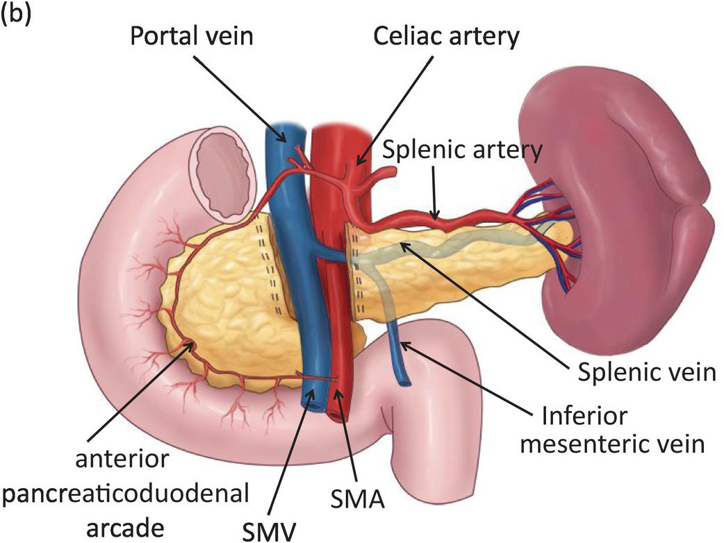
Compared to the size of the organ, the splenic artery has a large diameter. Near the gate, it splits into 6 - 8 branches, each included separately in the thickness of the organ, where they give small branches, grouping in the form of tassels, penicilli .
Arterial capillaries pass into the venous sinuses, the walls of which are formed by endothelial syncytium with numerous gaps through which blood elements enter the venous sinuses. The venous trunks starting from here, in contrast to arterial ones, form numerous anastomoses among themselves. The roots of the splenic vein (veins of the 1st order) carry blood from relatively isolated areas of the organ parenchyma, called the spleen zones.
A zone means a part of the intraorgan venous bed of the spleen, which corresponds to the distribution of the 1st order vein. The zone occupies the entire diameter of the organ. In addition to zones, segments are also distinguished.
The segment is a 2nd order vein distribution pool; it forms part of the zone and is located, as a rule, on one side of the hilum of the spleen.
The number of segments varies within a wide range - from 5 to 17. Most often, the venous bed consists of 8 segments. Depending on the position in the organ, they can be designated as anterior pole segment, anterior superior, anterior inferior, middle superior, middle inferior, posterior superior, posterior inferior and posterior pole segment.
The splenic vein flows into v. portae. The pulp does not contain lymphatic vessels. The nerves from the plexus coeliacu s penetrate along with the splenic artery.
Development. The spleen is laid in the mesogastrium posterius in the form of an accumulation of mesenchymal cells at the 5th week of uterine life. In newborns, the spleen is relatively bulky (1 - 15 g). After 40 years, a gradual decrease in the spleen is noticeable.
Video lesson topography of the spleen, its blood supply and innervation


Comments
Post a Comment